16200 Metcalf Ave, Overland Park, KS 66085

Pet Ultrasound or X-Ray: Which Scan Helps Find Common Health Problems?
By Blue Valley Animal Hospital | September 25, 2026
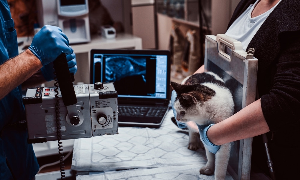
When a dog or cat develops unexplained vomiting, abdominal pain, breathing changes, limping, weight loss, or other concerning symptoms, diagnostic imaging can help a veterinarian look beyond what can be detected during a physical examination. X-rays and ultrasound are two commonly used imaging methods, but they provide different types of information. The right scan depends on the symptoms, body area being evaluated, and suspected health problems.
At Blue Valley Animal Hospital, diagnostic imaging can be considered alongside a pet’s history, physical examination, and other testing to help determine what may be causing unusual symptoms. Understanding the differences between X-rays and pet ultrasound can help owners know why a veterinarian may recommend one test over the other, or sometimes both.
What Is an X-Ray for Pets?
An X-ray, also called a radiograph, uses a small amount of radiation to create images of structures inside the body. It is particularly useful for evaluating bones, the chest, and the overall size and position of certain organs.
X-rays can help veterinarians investigate conditions such as:
- Bone fractures
- Arthritis and other skeletal changes
- Certain joint problems
- Chest abnormalities
- Some lung conditions
- Heart size and shape
- Certain abdominal abnormalities
- Swallowed foreign objects
- Abnormal gas patterns
X-rays are especially useful when a veterinarian needs a quick overview of a particular area of the body.
What Is a Pet Ultrasound?
An ultrasound uses sound waves to create real-time images of internal structures. Unlike X-rays, ultrasound does not use ionizing radiation.
Ultrasound can provide detailed information about soft tissues and can show movement within the body. It is commonly used to evaluate abdominal organs such as the liver, kidneys, spleen, bladder, and gastrointestinal tract.
Depending on the clinical situation, ultrasound may help investigate:
- Abdominal masses
- Changes in internal organs
- Certain gastrointestinal problems
- Urinary tract concerns
- Liver or gallbladder abnormalities
- Reproductive system conditions
- Fluid within the abdomen
- Some soft-tissue abnormalities
Because ultrasound produces real-time images, it can provide information about the structure and appearance of organs that may be difficult to assess with standard X-rays.
Pet Ultrasound vs. X-Ray: What’s the Difference?
The two imaging methods complement each other rather than competing directly.
| Feature | X-Ray | Ultrasound |
| Uses radiation | Yes | No |
| Best for | Bones, chest, overall structures | Soft tissues and internal organs |
| Real-time imaging | No | Yes |
| Bone detail | Excellent | Limited |
| Soft-tissue detail | More limited | Often better |
| Foreign objects | Can detect some | May help identify some |
| Abdominal evaluation | Useful | Provides more detailed soft-tissue assessment |
The veterinarian chooses imaging based on the clinical question rather than simply selecting whichever scan is considered more advanced.
When Is an X-Ray Usually Recommended?
An X-ray may be useful when a pet has symptoms involving the bones, chest, or certain abdominal concerns.
Suspected Fractures
After an accident, fall, or other trauma, X-rays can help identify broken bones and provide information about the location and severity of an injury.
Breathing Problems
Chest radiographs can help evaluate the lungs, heart silhouette, and other structures within the chest. They may be recommended when a pet has coughing, difficulty breathing, or other respiratory symptoms.
Swallowed Objects
Some swallowed objects are visible on X-rays. Others may not be directly visible, but changes in the position or appearance of the gastrointestinal tract can still provide useful information.
If a pet has swallowed an object, prompt veterinary evaluation is important because the appropriate diagnostic approach depends on what was swallowed and where it may be located.
When Is Ultrasound Usually Recommended?
Ultrasound is particularly valuable when the veterinarian needs a closer look at soft tissues or abdominal organs.
Abdominal Problems
Pets with unexplained vomiting, diarrhea, abdominal discomfort, appetite changes, or weight loss may require abdominal imaging. Ultrasound can provide detailed views of several organs and may help identify abnormalities that are not easily characterized with X-rays alone.
Suspected Masses
If another examination or imaging test identifies an abnormal structure, ultrasound may help determine its location, size, internal appearance, and relationship to nearby tissues.
In some cases, ultrasound can also help guide a needle toward a specific area for sampling when tissue or fluid evaluation is appropriate.
Urinary Concerns
The bladder and parts of the urinary system can be examined using ultrasound. It may help identify certain bladder abnormalities, stones, thickening, or other changes.
Can a Pet Need Both an X-Ray and an Ultrasound?
Yes. In some situations, using both imaging techniques provides more complete information.
For example, an X-ray may first provide an overview of the abdomen and show an unusual gas pattern or possible foreign material. Ultrasound may then provide more detailed information about the affected organs and surrounding soft tissues.
Similarly, chest X-rays and other diagnostic tests may be used together when investigating complex respiratory or cardiovascular concerns.
The goal is not to perform unnecessary tests. Instead, the veterinarian considers which diagnostic information is most likely to answer the clinical question.
Does My Pet Need Anesthesia for Imaging?
Not every pet requires anesthesia for an X-ray or ultrasound.
Many pets can undergo imaging while awake if they are calm and can remain safely positioned. Sedation may sometimes be recommended when a pet is extremely anxious, painful, difficult to position, or unable to remain still.
Anesthesia or sedation decisions depend on the procedure, the pet’s health, temperament, and the type of imaging being performed.
How Veterinarians Decide Which Scan to Use
The decision begins with a thorough history and physical examination.
A veterinarian may consider:
- Your pet’s age
- Breed and size
- Current symptoms
- Duration of symptoms
- Previous medical history
- Recent injuries
- Appetite and weight changes
- Laboratory test results
- Findings from earlier imaging
For example, a limping dog after trauma may need an X-ray to evaluate the bones and joints, while a cat with unexplained weight loss and abdominal symptoms may benefit from an ultrasound.
The appropriate imaging method is therefore based on the problem being investigated.
What Should Pet Owners Expect During Imaging?
Before imaging, the veterinary team may provide instructions regarding food, water, medications, or activity, depending on the planned procedure.
For an abdominal ultrasound, fasting may sometimes be recommended because food and gas in the digestive tract can interfere with visualization. The abdomen may also need to be clipped to allow the ultrasound probe to make proper contact with the skin.
For X-rays, positioning is important because movement can blur images. The veterinary team will take appropriate safety precautions during the procedure.
After imaging, the veterinarian reviews the findings and explains what they may mean. Additional testing may be recommended if the images identify an abnormality that requires further investigation.
When Should You Ask a Veterinarian About Diagnostic Imaging?
Imaging may be appropriate when symptoms persist, recur, worsen, or cannot be explained through a physical examination and routine testing alone.
Contact a veterinarian if your pet develops concerning signs such as:
- Persistent vomiting
- Unexplained weight loss
- Abdominal pain or swelling
- Difficulty breathing
- Sudden lameness
- Significant injury
- Loss of appetite
- Repeated urinary problems
- Suspected foreign-object ingestion
- Unexplained weakness
Sudden or severe symptoms may require prompt veterinary evaluation rather than waiting for a routine appointment.
Supporting Better Diagnostic Decisions
X-rays and ultrasound each have important strengths. X-rays are particularly valuable for bones, chest structures, and certain foreign objects, while ultrasound provides detailed, real-time information about many soft tissues and abdominal organs.
Neither test is automatically better for every condition. The most appropriate choice depends on what the veterinarian needs to investigate.
For pet owners in Overland Park, KS, schedule your appointment with Blue Valley Animal Hospital if your pet develops unexplained symptoms that may require diagnostic imaging. A veterinary evaluation can help determine which diagnostic approach is appropriate for your pet.
FAQs
Q1. Is an X-ray or ultrasound better for pets?
Ans: Neither is universally better. X-rays are particularly useful for bones, the chest, and certain foreign objects, while ultrasound generally provides more detailed information about soft tissues and many abdominal organs. The appropriate test depends on the suspected problem.
Q2. Is ultrasound safe for dogs and cats?
Ans: Ultrasound does not use ionizing radiation and is generally considered a safe diagnostic imaging method. The veterinary team will determine whether it is appropriate based on the pet’s symptoms and overall health.
Q3. Can an X-ray show a swallowed foreign object?
Ans: Some foreign objects can be seen directly on X-rays, while others cannot. Even when an object is not visible, an X-ray may reveal changes that suggest an obstruction or other gastrointestinal problem.
Q4. Does my pet need to be sedated for an ultrasound?
Ans: Not always. Many pets can undergo ultrasound while awake. Sedation may be considered if the pet is highly anxious, painful, unable to remain still, or requires a procedure that involves sampling tissue or fluid.
Q5. Why would my pet need both an X-ray and ultrasound?
Ans: The two tests provide different information. An X-ray can offer an overview of structures such as bones and the chest, while ultrasound can provide detailed views of soft tissues and organs. Using both may help investigate a complicated condition more thoroughly.
Q6. Should I wait if my pet has unexplained vomiting or abdominal pain?
Ans: Persistent vomiting or abdominal pain should be discussed with a veterinarian, particularly if accompanied by weakness, loss of appetite, abdominal swelling, repeated vomiting, or other concerning symptoms. A veterinary examination can determine whether diagnostic imaging is needed.